dicomreadVolume
Create 4-D volume from set of DICOM images
Syntax
Description
V = dicomreadVolume(sourceTable)sourceTable. The table must contain only one row that
specifies the metadata for a DICOM volume.
V = dicomreadVolume(sourceTable,rowname)rowname of the multirow table. Use this syntax when
sourceTable contains multiple rows.
Examples
Load volume data from a folder containing DICOM image files. Use the squeeze function to remove any singleton dimensions.
[V,spatial,dim] = dicomreadVolume("dog");
V = squeeze(V);
Display the 4-D DICOM volume. Generate a colormap and transparency map for magnetic resonance (MR) images.
intensity = [0 20 40 120 220 1024];
alpha = [0 0 0.15 0.3 0.38 0.5];
color = ([0 0 0; 43 0 0; 103 37 20; 199 155 97; 216 213 201; 255 255 255])/ 255;
queryPoints = linspace(min(intensity),max(intensity),256);
amap = interp1(intensity,alpha,queryPoints)';
cmap = interp1(intensity,color,queryPoints);
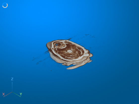
View the volume with the custom colormap and transparency map.
volshow(V,Colormap=cmap,Alphamap=amap);
Display the returned spatial structure from dicomreadVolume. The structure contains spatial information about the input DICOM image files.
spatial
spatial = struct with fields:
       PatientPositions: [22×3 double]
          PixelSpacings: [22×2 double]
    PatientOrientations: [2×3×22 double]
              ImageSize: [512 512 22]
Display the dimension information from dicomreadVolume. The value specifies that the slice offset is largest along the z-dimension.
dim
dim = 3
Gather details about the DICOM files contained in a folder by using the dicomCollection function. The function returns the details of the available DICOM metadata in the form of a table.
sourceTable = dicomCollection("dog");Display the table. Each row contains the metadata for the a DICOM image set present in the specified folder.
sourceTable
sourceTable=1×14 table
             StudyDateTime           SeriesDateTime         PatientName      PatientSex    Modality    Rows    Columns    Channels    Frames    StudyDescription    SeriesDescription                        StudyInstanceUID                                             SeriesInstanceUID                          Filenames
          ____________________    ____________________    _______________    __________    ________    ____    _______    ________    ______    ________________    _________________    _________________________________________________________    _________________________________________________________    _____________

    s1    14-Dec-2013 15:47:31    14-Dec-2013 15:54:33    "GORBERG MITZI"       "F"          "MR"      512       512         1          22           "CSP"               "AX T2"         "1.2.840.113619.2.244.3596.11880862.13689.1386517653.214"    "1.2.840.113619.2.244.3596.11880862.13689.1386517653.217"    {22×1 string}
Create a 4-D DICOM volume from a DICOM image set in the table. Specify the row name that contains the desired DICOM image set. To create an isotropic volume, specify the MakeIsotropic name-value argument as true. Use the squeeze function to remove any singleton dimensions.
V = dicomreadVolume(sourceTable,"s1",MakeIsotropic=true);
V = squeeze(V);
Generate a colormap and transparency map for MRI images.
intensity = [0 20 40 120 220 1024];
alpha = [0 0 0.15 0.3 0.38 0.5];
color = ([0 0 0; 43 0 0; 103 37 20; 199 155 97; 216 213 201; 255 255 255])/255;
queryPoints = linspace(min(intensity),max(intensity),256);
amap = interp1(intensity,alpha,queryPoints)';
cmap = interp1(intensity,color,queryPoints);
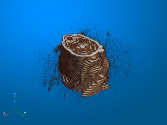
Display the isotropic 4-D DICOM volume with the custom colormap and transparency map by using the volshow function.
vol = volshow(V,Colormap=cmap,Alphamap=amap);
Input Arguments
Output Arguments
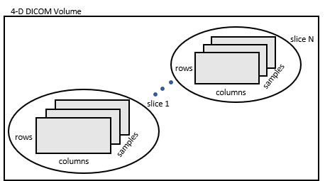
4-D DICOM volume, returned as a numeric array.
The dimensions of V are [rows,
columns, samples,
slices], where samples is the
number of color channels per voxel. For example, grayscale volumes have one
sample, and RGB volumes have three samples. Use the squeeze function to remove
any singleton dimensions, such as when the sample is 1.
Location, resolution, and orientation of slices collected from the metadata of the input DICOM images, returned as a structure with these fields.
| Field | Description |
|---|---|
PatientPositions | (x, y, z) coordinate of the first pixel in each slice, measured in millimeters from the origin of the scanner coordinate system. |
PixelSpacings | Distance between neighboring rows and columns within each slice, in millimeters. |
PatientOrientations | Pair of direction-cosine triplets that designate the direction of the rows and columns in each slice relative to the patient position. |
For more information about DICOM attributes, see part 3 of the DICOM standard, section C.7.6.2.
Dimension with the largest offset, returned as 1,
2, or 3. The value denotes the
dimension in a 3-D coordinate system that has the largest amount of offset
between adjacent slices in the input DICOM data.
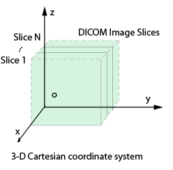
If the largest offset is along the x dimension, then
dimis1.If the largest offset is along the y dimension, then
dimis2.If the largest offset is along the z dimension, then
dimis3.
Extended Capabilities
Version History
Introduced in R2017bSee Also
DICOM Browser | dicominfo | dicomread | dicomCollection | tiffreadVolume